Hydrogels and Graphene: The Technological Fusion Revolutionizing Materials Science 

Hydrogels and Graphene:

The Technological Fusion Revolutionizing Materials Science 

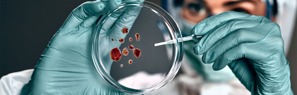
Hydrogels are polymeric networks with a hydrophilic structure that allows them to retain large amounts of water in their three-dimensional networks. From their first references in 1900 to the advancements by Wichterle and Lim in the 1960s, hydrogel technology has evolved to become a vital solution in fields such as medicine (for controlled drug or bioactive agent delivery), environmental remediation (for contaminant adsorption or soil restoration), agriculture (for water retention or soil conditioning), food industry (from texturizing agents to smart packaging), and even energy storage, among others.  

“In 1900, the term ‘hydrogel’ first appeared in scientific literature to describe a colloidal gel of inorganic salts.” 

Hydrogels may be chemically crosslinked through covalent bonds, physically through non-covalent interactions, or via a combination of both. They are classified into natural (including proteins such as collagen and gelatin, and polysaccharides such as starch, alginate, and agarose), synthetic (produced through chemical polymerization methods), and hybrid hydrogels. Synthetic materials have largely replaced natural ones due to better water absorption, longer lifespan, and a broader variety of raw materials.  

“The water absorption capacity of hydrogels is due to hydrophilic functional groups attached to their polymer structure, while their resistance to dissolution results from crosslinks between the network chains.” 

Hydrogels are mainly known and used for their excellent water absorption capabilities without altering their structure. However, their low mechanical strength and sensitivity to external stimuli such as temperature, light, or electric fields can either enhance or limit their performance in dynamic environments. Some examples include: 

Temperature Sensitivity: These hydrogels expand or contract in response to heat or cold, enabling them to release contaminants, drugs, or bioactive agents—ideal for wastewater treatment or medical therapies. Their challenges include long-term stability and precise temperature responsiveness. 

Photosensitivity: The integration of photoactive agents into their polymer networks can improve contaminant degradation, activate and release specific drugs, or enhance cellular growth conditions in tissue engineering. However, continuous UV exposure may lead to photodegradation of the material. 

Electric Field Sensitivity: Electroactive hydrogels incorporate ionic groups that move and alter the hydrogel’s structure under an electric field, modifying its permeability to allow or block the passage of substances. This is useful for wastewater treatment and controlled water or nutrient release in agriculture. High costs and limited durability remain key challenges. 

pH Sensitivity: This is achieved by incorporating ionizable functional groups (carboxyl or amino), which gain or lose protons based on environmental pH, triggering structural changes that allow water absorption or release. 

How Does Nanotechnology Enhance Hydrogel Performance?  

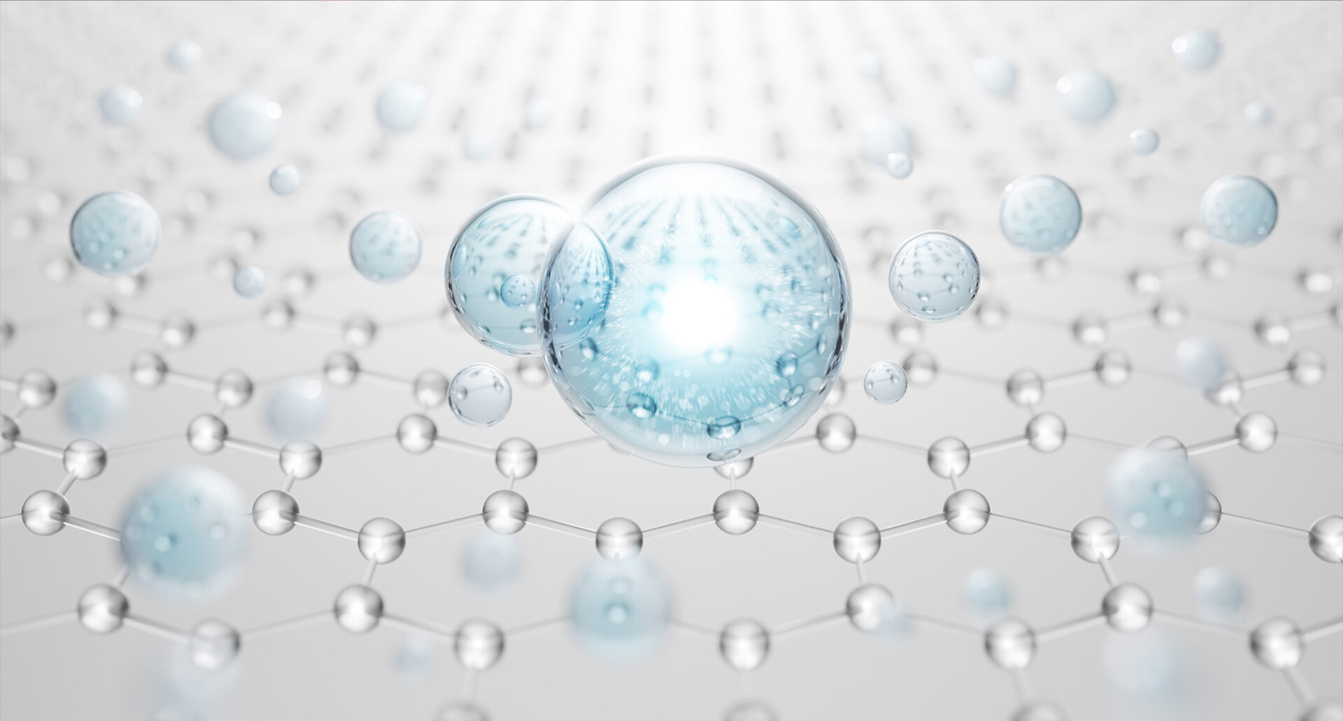
Nanotechnology—the interdisciplinary field focused on manipulating and manufacturing materials at the atomic and molecular scale (1–100 nanometers)—has made significant contributions to hydrogels, particularly in improving mechanical strength and developing smart functionalities for biomedical and environmental applications.  

“To understand the nanoscale, consider that the average thickness of a human hair is approximately 60,000 nm, while a nanometer is one-millionth of a millimeter.” 

What Added Value Does Graphene Bring to Hydrogels?  

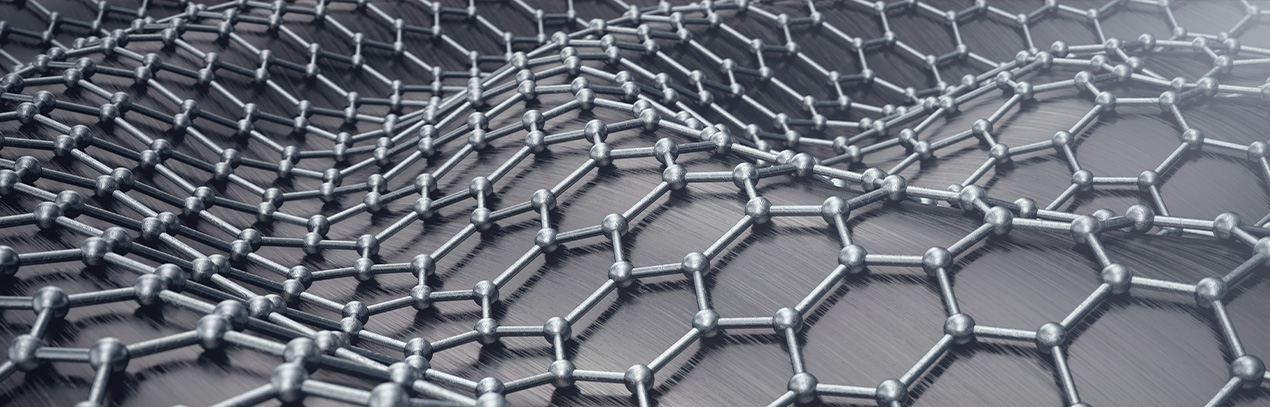
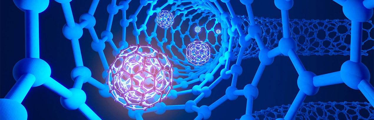
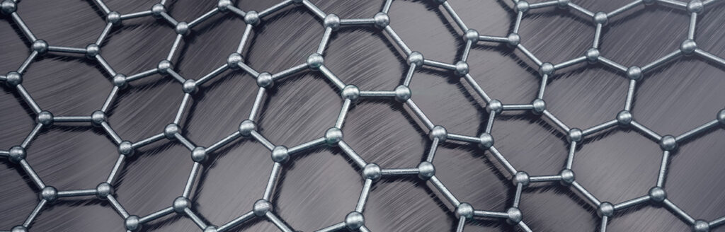
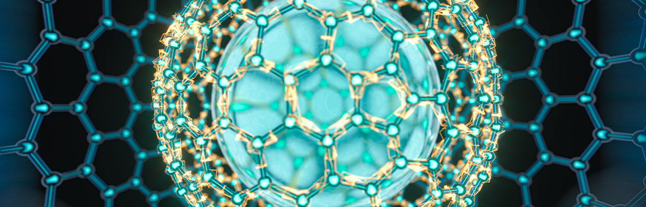
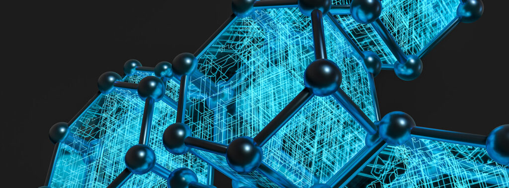
Graphene is a nanometric, two-dimensional (2D) carbon sheet one atom thick, with a structure similar to a benzene ring. It has attracted significant research interest due to its exceptional properties: thermal conductivity, mechanical strength, flexibility, and biocompatibility, among others, which can be transferred to hydrogel matrices. 

1. Mechanical Strength: 

One of the key limitations of hydrogels is their low mechanical resistance, making them unsuitable for high-stress environments. Graphene can significantly reinforce hydrogel structure, improving durability and mechanical stability. 

2. Thermal Stability: 

Hydrogels are sensitive to temperature changes, which can affect their performance. Graphene can enhance their thermal stability, maintaining functional properties across wider temperature ranges. 

3. Antimicrobial Protection and Biocompatibility: 

While hydrogels are generally biocompatible, scientific evidence shows that adding graphene improves their stability and compatibility. Its intrinsic antimicrobial properties also make hydrogels safer for infection-sensitive applications. 

4. Electrical Responsiveness: 

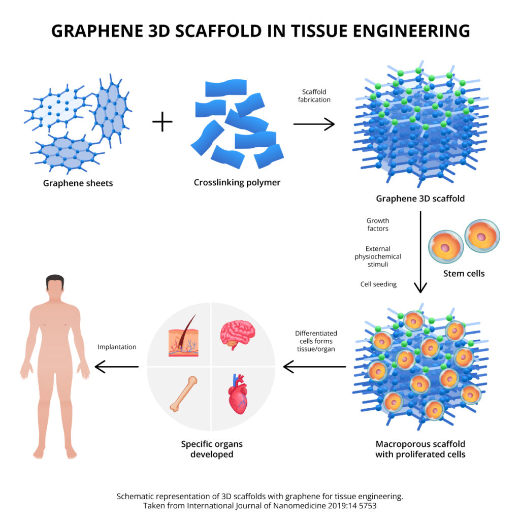
Graphene’s ability to transmit electrical signals supports cell communication in electrically active tissues (e.g., nerve, muscle, heart). It also enables controlled drug release under voltage stimulation, while minimizing overheating during electrical activation. 

Although there are no graphene-based hydrogels commercially available yet, many experimental nanotechnological developments around the world are laying the groundwork for future applications, including: 

  1. Spain (2025): A study by the Spanish National Research Council and the National Hospital for Paraplegics used a reduced graphene oxide (rGO) foam scaffold to reconnect severed spinal cords in rats, promoting neuronal reconnection and vascular regeneration. 
  1. Argentina (2025): The University of Buenos Aires, CONICET, and INTI developed hydrogels resistant to hydration-dehydration cycles for nanofiltration of viruses, bacteria, fungi, and heavy metals from water. 
  1. USA (2023): A study published in Environmental Science & Technology demonstrated that graphene-based hydrogels could remove up to 95% of lead ions from aqueous solutions. 
  1. China (2022): Biomaterials Translational published a study on a hyaluronic acid–graphene oxide hydrogel combined with Senexin A for treating vascular occlusive diseases, achieving sustained release over 21 days and good biocompatibility. 
  1. USA (2022): In Applied Materials & Interfaces, researchers reported a graphene hydrogel scaffold that promoted cartilage regeneration with type II collagen expression and stable cellular growth. 
  1. Spain (2018): Researchers at the University of the Basque Country developed a starch–graphene hydrogel for flexible brain implant electrodes. The graphene was stabilized in water using sage extract, which also added antibacterial and electrical properties. 
  1. USA (2017): A porous graphene oxide hydrogel developed for water purification showed enhanced contaminant adsorption due to improved stability, nanotransport channels, and hydrogen bonding. 
  1. USA (2017): A soft, injectable hydrogel made from PEGDA–melamine and GO improved cardiac function in rats with myocardial infarction, reducing infarct size and fibrosis while promoting neovascularization. 

Conclusion 

Graphene-based hydrogels, and those incorporating its derivatives, offer significant structural and functional improvements. Their development represents a leap toward smarter and more adaptive systems in biomedicine, agriculture, and environmental remediation. However, like any emerging technology, widespread adoption will require overcoming regulatory and large-scale manufacturing challenges.  

Written by: EF/ DHS 

References 

  1. Visan, A. I.; Negut, I. Environmental and Wastewater Treatment Applications of StimulusResponsive Hydrogels. Gels 2025, 11 (1), 72.  
  1. Yu, K.; Wang, D.; Wang, Q. Tough and SelfHealable Nanocomposite Hydrogels for Repeatable Water Treatment. Polymers 2018, 10, 880. 
  1. Lim, S. L.; Tang, W. N. H.; Ooi, C. W.; Chan, E.S.; Tey, B. T. Rapid swelling and deswelling of semiinterpenetrating network poly(acrylic acid)/poly(aspartic acid) hydrogels prepared by freezing polymerization. J. Appl. Polym. Sci. 2016, 133, 9. 
  1. Yuan, Z.; Wang, Y.; Han, X.; Chen, D. The adsorption behaviors of the multiple stimulusresponsive poly(ethylene glycol)based hydrogels for removal of RhB dye. J. Appl. Polym. Sci. 2015, 132, 42244. 
  1. Thakur, S.; Arotiba, O. Synthesis, characterization and adsorption studies of an acrylic acidgrafted sodium alginatebased TiO₂ hydrogel nanocomposite. Adsorpt. Sci. Technol. 2018, 36, 458–477. 
  1. Eraković, Z.; Stefanović, D. Purification of contaminated wastewater with the help of graphene composites with hydrogels. Facta Univ. Ser. Work. Living Environ. Prot. 2022, 19, 27–**. 
  1. Wu, R.; Tian, L.; Wang, W.; Man, X. Bifunctional cellulose derivatives for the removal of heavymetal ions and phenols: Synthesis and adsorption studies. J. Appl. Polym. Sci. 2015, 132, 41830. 
  1. Zheng, Y.; Zhu, Y.; Wang, F.; Wang, A. GelatinGrafted Granular Composite Hydrogel for Selective Removal of Malachite Green. Water, Air, Soil Pollut. 2015, 226, 354. 
  1. Malik, R.; Saxena, R.; Warkar, S. G. Organic Hybrid Hydrogels: A Sustenance Technique in WasteWater Treatment. ChemistrySelect 2023, 8, e202203670. 
  1. Berg, J.; Seiffert, S. Composite hydrogels based on calcium alginate and polyethyleneimine for wastewater treatment. J. Polym. Sci. 2023, 61, 2203. 
  1. Singh, R.; Datta, B. Advances in Biomedical and Environmental Applications of Magnetic Hydrogels. ACS Appl. Polym. Mater. 2023, 5, 5474. 
  1. Tang, S. C. N.; Yan, D. Y. S.; Lo, I. M. C. Sustainable Wastewater Treatment Using Microsized Magnetic Hydrogel with Magnetic Separation Technology. Ind. Eng. Chem. Res. 2014, 53, 15718. 
  1. Wahid, F.; Zhao, X.J.; Jia, S.R.; Bai, H.; Zhong, C. Nanocomposite hydrogels as multifunctional systems for biomedical applications: Current state and perspectives. Composites Part B: Engineering 2020, 200, 108208. 
  1. Bao, R.; Tan, B.; Liang, S.; Zhang, N.; Wang, W.; Liu, W. A ππ conjugationcontaining soft and conductive injectable polymer hydrogel highly efficiently rebuilds cardiac function after myocardial infarction. Biomaterials 2017, 122, 63. 
  1. Maturavongsadit, P.; Wu, W.; Fan, J.; Roninson, I. B.; Cui, T.; Wang, Q. Grapheneincorporated hyaluronic acidbased hydrogel as a controlled Senexin A delivery system. Biomater. Transl. 2022, 3 (2), 152–161.  
  1. Lyu, C.; Cheng, C.; He, Y.; Qiu, L.; He, Z.; Zou, D.; Li, D.; Lu, J. Graphene hydrogel as a porous scaffold for cartilage regeneration. ACS Appl. Mater. Interfaces 2022, 14 (49), 54431. 
  1. T. Sawyer, Principles of Nanotechnology, Murphy & Moore Publishing, 2022. 
  1. https://www.infocampo.com.ar/hidrogeles-con-nanotecnologia-un-desarrollo-cientifico-argentino-que-mejora-la-calidad-del-agua/ 
  1. https://www.agenciasinc.es/Noticias/Hidrogel-de-grafeno-y-almidon-para-electrodos-de-implantes-cerebrales 
  1. https://cadenaser.com/nacional/2025/01/30/cientificos-del-csic-logran-reconectar-la-medula-espinal-totalmente-seccionada-de-una-rata-cadena-ser 

Biocompatibility and Biodegradability of Graphene: Advances and Scientific Evidence

Biocompatibility and Biodegradability of Graphene:

Advances and Scientific Evidence

Graphene is widely recognized for its exceptional properties and its potential to revolutionize various industries. However, as a relatively recent technology with emerging applications, concerns have arisen regarding its impact on human health and the environment. Therefore, it is essential to analyze scientific studies that have evaluated its biocompatibility and biodegradability, providing evidence of its safety and behavior in different biological systems.

Biocompatibility: Defined as the absence of allergic or immune adverse reactions to a material in the body

Over the past decade, multiple studies have demonstrated that graphene and its derivatives can be biocompatible under certain conditions. Research on its interaction with blood, cell differentiation, kidney function, neuronal activation, and bone regeneration has yielded positive results. The following key findings stand out:

2012 – Compatibility with blood and macrophage response. The nanotoxicity of graphene on macrophages was evaluated based on its effects on metabolic activity, membrane integrity, oxidative stress induction, hemolysis, platelet activation and aggregation, coagulation cascade, cytokine induction, and immune cell activation and suppression.

Results indicated that while graphene does interact with macrophages, toxicity is significantly reduced through surface functionalization. Regarding blood compatibility, both functionalized and non-functionalized graphene exhibited excellent compatibility with red blood cells, platelets, and plasma coagulation pathways, with minimal alteration in cytokine expression by human peripheral blood mononuclear cells. Additionally, no premature immune cell activation or suppression was observed up to a relatively high concentration of graphene (75 μg mL⁻¹) after 72 hours of in vitro incubation.

Conclusion: Possible graphene toxicity can be easily avoided through surface functionalization.

A. Sasidharan, et. al., Hemocompatibility and Macrophage Response of Pristine and Functionalized Graphene, Small, 2012, 8, 1251

2014- Cardiac cell differentiation. The effect of graphene on the cardiomyogenic differentiation of human embryonic stem cells (hESCs) was analyzed. Graphene was synthesized via CVD and deposited on vitronectin-coated glass, a multifunctional protein found in plasma, platelets, and the extracellular matrix, to ensure hESC viability. Cells were cultured for 21 days, and results showed that graphene promoted the expression of genes involved in gradual differentiation into mesodermal and endodermal lineage cells and subsequently into cardiomyocytes, compared to cultures on glass without graphene.

Conclusion: Graphene can provide a platform for developing stem cell therapies for heart diseases by enhancing the cardiomyogenic differentiation of human embryonic stem cells.

Tae-Jin Lee, et. al., Graphene enhances the cardiomyogenic differentiation of human embryonic stem cells, Biochem Biophys Res Commun, 2014, 452(1):174

2016- Impact on kidney function. The effect of intravenously administered graphene oxide (GO) on the kidneys of mice was studied. Results showed that GO was excreted through urine, indicating rapid transit through the glomerular filtration barrier (GFB) without nephrotoxicity. The analysis concluded an absence of kidney function impairment up to one month after GO injection at increasing doses. Histological examination found no damage to the glomerular and tubular regions of the kidneys. Ultrastructural analysis also revealed no damage or changes in podocyte slit size, endothelial cell fenestra, or glomerular basement membrane width. Endothelial and podocyte cultures restored their barrier function after >48 hours of GO exposure, with significant cellular uptake observed in both cell types after 24 hours.

Conclusion: GO is not toxic to the kidneys..

Dhifaf A. Jasim, et. al., The Effects of Extensive Glomerular Filtration of Thin Graphene Oxide Sheets on Kidney Physiology. ACS Nano 2016, 10, 12, 10753

2018- Effect on Neuronal Activation. The effect of monolayer graphene on neuronal activation was evaluated. It was identified that graphene modifies membrane-associated functions in cultured cells, meaning it adjusts the distribution of extracellular ions at the interface with neurons—a key regulator of neuronal excitability.

The observed membrane changes included stronger potassium ion currents and a shift in the fraction of neuronal activation phenotypes from adaptive to tonic activation. The study’s hypothesis suggested that graphene-ion interactions are maximized when single-layer graphene is deposited on electrically insulating substrates.

Conclusion: Graphene oxide can act as a substrate for neuronal interaction.

N. P. Pampaloni, et. al., Single-layer graphene modulates neuronal communication and augments membrane ion currents, Nat. Nanotechnol., 2018, 13, 755

2018- Adjuvant in the Proliferation of Pulmonary and Neuronal Cells. Graphene oxide “papers” of different sizes and thicknesses were fabricated as a substrate for the culture of human pulmonary and neuronal cells. Their capacity for cell adhesion and proliferation was evaluated, along with a possible cytotoxic response by detecting lactate dehydrogenase (LDH) in cell supernatants.

Conclusion: Graphene oxide can act as a biocompatible cellular substrate for cell growth without cytotoxic effects, opening greater possibilities for tissue engineering, regenerative medicine, and bionic applications.D. A. Jasim, et. al., Graphene-based papers as substrates for cell growth: Characterisation and impact on mammalian cells, FlatChem, 2018, 12, 17

2020- Biocompatibility of Graphene in Dental Materials. The biocompatibility of a graphene-containing restorative material and dental cement was studied on a mandibular defect in an animal model. Cytotoxicity was evaluated in vitro at 24 hours on human dental follicle stem cells and oral keratinocytes. In vivo studies were conducted seven weeks after implantation, including histological analysis of collected bone tissue, plasma biochemistry, oxidative stress assessment, and subchronic organ toxicity analysis.

The in vitro results showed that the materials did not induce toxicity in cells. In vivo, the animal models exhibited no symptoms of acute toxicity or local inflammation. No changes were detected in organ weights, and histological analysis revealed no alterations in liver or kidney tissues. Systemic toxicity of the materials in organs was not observed.

Conclusion: The study provides further evidence of the potential of graphene-based dental materials for bone regeneration and biocompatibility.

A. Dreanca, et. al., Systemic and Local Biocompatibility Assessment of Graphene Composite Dental Materials in Experimental Mandibular Bone Defect. Materials 2020, 13, 2511; doi:10.3390/ma13112511

2022- Risks of Graphene in Microplastics. The study was conducted on a composite of polyamide 6 or Nylon-6, a plastic commonly used in the automotive and sports industries, reinforced with reduced graphene oxide (rGO 2.5%). The material was then subjected to wear to emulate natural processes throughout its useful life, releasing particles approximately between 1.9 µm and 3.2 µm in size. To analyze the effects of the worn particles along the most likely exposure routes, in vitro human cell models of the lungs, gastrointestinal tract, skin, and immune system were used, as well as an animal model to study pulmonary exposure in vivo.

At the end of the study, only limited acute responses were found after exposure to the microplastics in the different models. Only the free rGO induced significant adverse effects, particularly in macrophages.

Conclusion: Microplastics with graphene suggest a low risk to human health. Graphene materials should not be inhaled.

S. Chortarea, et. al., Hazard assessment of abraded thermoplastic composites reinforced with reduced graphene oxide, Journal of Hazardous Materials 435 (2022) 129053.

2023- Pulmonary Function. The biological response, distribution, and biopersistence of four types of graphene in the lungs of mice were analyzed up to 28 days after a single oropharyngeal aspiration. The results showed that none of the materials induced a strong pulmonary immune response, with neutrophils being more effective at internalizing, degrading, and eliminating small graphene sheets (~50nm) than macrophages, as larger sheets (~8µm) may have greater persistence.

Conclusion: Graphene does not cause an inflammatory response in the lungs; however, it is important to consider the size of the sheets, as smaller ones are easier to eliminate from the airways and, therefore, safer.

Thomas Loret, et. al., Lung Persistence, Biodegradation, and Elimination of Graphene-Based Materials are Predominantly Size-Dependent and Mediated by Alveolar Phagocytes, Small, 2023,19(39): e2301201

2024- Pulmonary and Cardiovascular Function. An in vivo study was conducted to evaluate the effect of graphene oxide inhalation on pulmonary and cardiovascular function in healthy humans. For the trial, 14 volunteers inhaled small and ultrafine graphene oxide sheets at a controlled concentration during two-hour repeated visits. Heart rate, blood pressure, pulmonary function, and inflammatory markers were unaffected regardless of particle size; blood analysis showed few differential plasma proteins, and thrombus formation increased slightly in an ex vivo arterial injury model.

Conclusion: Graphene oxide inhalation can be tolerated and is not associated with apparent harmful effects in healthy humans. The study lays the groundwork for further human studies that examine a larger number of individuals as well as different types and doses of graphene.

Jack P. M. Andrews, First-in-human controlled inhalation of thin graphene oxide nanosheets to study acute cardiorespiratory responses. Nature nanotechonoly, 2024, 19, 705.

Biodegradation: Process by which a substance is broken down by living organisms through enzymatic or metabolic mechanisms

One of the most relevant aspects in evaluating the safety of graphene is its biodegradability. Research conducted under the Graphene Flagship project has demonstrated that graphene and graphene oxide can be successfully degraded, as follows:

2018 – Researchers affiliated with the European Union’s Graphene Flagship project, from institutions such as the National Center for Scientific Research (CNRS) in France, the University of Strasbourg, the Karolinska Institute, and the University of Castilla-La Mancha (UCLM), through studies such as “Dispersibility-Dependent Biodegradation of Graphene Oxide by Myeloperoxidase” (2015), “Graphene Oxide is Degraded by Neutrophils and the Degradation Products Are Non-Genotoxic” (2018), and “Peroxidase Mimicking DNAzymes Degrade Graphene Oxide” (2018), discovered that the enzyme myeloperoxidase (MPO) successfully degrades both graphene and graphene oxide.

Myeloperoxidase (MPO): An enzyme released by neutrophils, cells responsible for eliminating any foreign body or bacteria entering the body, present in the lungs. When a foreign body or bacteria is detected, neutrophils surround it and secrete MPO to destroy the threat.

Professor Andrea C. Ferrari, Head of Science and Technology at Graphene Flagship and Chairman of its Management Panel, stated: “The report on a successful pathway for graphene biodegradation is a very important step in ensuring the safe use of this material in applications. The Graphene Flagship has placed research into the health and environmental effects of graphene at the center of its program from the beginning. These results strengthen our confidence in the potential of graphene for biomedical and technological innovations.”

https://graphene-flagship.eu/materials/news/biodegradable-graphene/#:~:text=Ferrari%2C%20Science%20and%20Technology%20Officer,our%20innovation%20and%20technology%20roadmap%22

Cristina Martın, et al., Biocompatibility and biodegradability of 2D materials: graphene and beyond, Chem. Commun., 2019, 55, 5540

This discovery is crucial, as it confirms that graphene is not an accumulative material in the human body or the environment. Instead, it can be naturally processed and eliminated, reducing the risks of long-term toxicity.

Conclusion

Graphene and its derivatives have demonstrated a high degree of biocompatibility and controlled degradability in various scientific studies. While challenges remain, current evidence supports its safety in biomedical, industrial, and environmental applications.

The key to its proper use lies in selecting the right type of graphene and its functionalization, which helps minimize risks and enhance its benefits. Thanks to advances in research, the viability of graphene as an innovative, safe, and sustainable material is becoming increasingly clear, with applications ranging from regenerative medicine to advanced nanotechnology.

The continuous development of scientific studies will further strengthen its position as one of the key technologies of the future, ensuring its responsible and effective implementation across different industries.

Authored by: EF/DHS

Graphene and nanomedicine: the perfect combination for improved health

Graphene and nanomedicine:

the perfect combination for improved health

Part III. Dentistry- Implantology

The application of nanotechnology in nanomedicine is based on the fact that most biological molecules, from DNA, amino acids and proteins to constituents such as hydroxyapatite and collagen fibrils, among others, exist and function at the nanometric scale.

Nanometer (nm): millionth of 1 millimeter.

Graphene materials are two-dimensional (2D) sheet-shaped carbon nanoparticles that have gained popularity in the field of biomedical sciences not only for their incredible mechanical, thermal, electrical, optical, and biological properties, but also for their ability to transfer these properties to other materials allowing the possibility of creating new compounds with advanced characteristics. In Odontology, and particularly in relation to implantology, this transfer of properties has opened numerous lines of research with great expectations due to the interesting synergistic effect between infection control and its regenerative capacity.1

Nanoparticle: particle that measures between 1 and 100 nm.

Graphene as a new strategy for the design and manipulation of dental implants and tissue regeneration. Taken from Tissue Eng Regen Med. 2017; 14(5):481

What are the problems that graphene could solve?

Osseointegration

One of the main concerns after the placement of an implant is the failure of its osseointegration. This can occur because instead of bone cells growing at the bone-implant interface, fibrous tissue grows that does not allow implant stabilization. An alternative to favor site conditions where cell interactions will occur is modification of the implant surface by physical or chemical methods to create nanoporosities that increase surface area and favor cell activity. 2

Osseointegration: Firm, stable, and long-lasting connection between an implant and the surrounding bone. Its success depends on biological and systemic factors of the patient, in addition to the characteristics of the implant.

In the case of graphene materials, in addition to their extensive and extremely thin surface area one atom thick, another of their added values is the cloud of electrons that surrounds them, and the presence of some oxygenated groups allows them to interact with proteins serum to form a focal adhesion. In other words, the hydrophobic/hydrophilic nature of these nanomaterials in combination with the roughness of the surface contributes to the interaction with proteins and later with cells, acting as a scaffold to promote the growth, differentiation, and anchorage of bone cells in the implant, paving the way for a stable and predictable osseointegration with a better projection of useful life.3,4

The regenerative impact of graphene materials lies in their great ability to adsorb proteins, creating a layer between cells and the surfaces of the materials to promote cell adhesion and proliferation.1

Infection control

Another cause for implant failure is the appearance of peri-prosthetic or peri-implant infections; to avoid them, it is common to use techniques such as antibiotic impregnation, local drug delivery systems, and the coating of implants with titanium nanotubes, silver nanoparticles, or polypeptide nanofilms for the controlled release of antibiotics.5 However, the worrying increase of antibiotic resistance has made these strategies less and less effective.

Graphene materials, in addition to their biocompatibility, have intrinsic antimicrobial properties with advantages over traditional antibiotics as they have less chance of developing microbial resistance. Odontology has been exploring these effects for several years on bioceramic materials such as alumina and zirconium, metals such as titanium, restorative materials such as glass ionomer, and polymeric materials such as polymethyl methacrylate (PMMA), to name a few. In general, the antimicrobial mechanisms accepted for these nanostructures are: 1) physical damage to the membrane, 2) oxidative stress, 3) inactivation by electron withdrawal, 4) isolation against the passage of nutrients and finally, 5) in the case of coatings, control of hydrophobicity and surface energy can also prevent cell attachment with low affinity and prevent biofilm formation.6,7

Biofilm: Layer of microorganisms that grow and adhere to the surface of a natural structure such as teeth (dental plaque) or artificial such as a medical device (intravascular catheters).

In 2021, a group of scientists from the University of Gwangju, Korea, published a study in which they coated zirconium implants with graphene oxide using the argon plasma method. Their results reported that this modification reduced by 58.5% the presence of Streptococcus mutans, the bacterium with the greatest influence on the formation of dental plaque and dental caries, agreeing with a significant reduction in biofilm thickness of 43.4%. In addition to the antimicrobial effect, they also showed a statistically significant increase of 3.2% and 15.7% in the proliferation and differentiation of bone cells.8 These results are consistent with what was reported by the Jiao Tong University, Shanghai, on a hybrid material of titanium with graphene. synthesized by the spark plasma sintering (SPS) technique. Similarly, the research demonstrated an interesting decrease in the formation of multibacterial biofilms composed of Streptococcus mutans, Fusobacterium nucleatum and Porphyromonas gingivalis, accompanied by an improvement in the activity of human gingival fibroblasts, one of the most important cell groups involved in healing.9 In addition to the synergy between infection control and its regenerative capacity, other studies related to dental implantology are also focusing their attention on the mechanical properties for the design of new implants or restorative materials. 10-12

Energeia-Graphenemex, the pioneering Mexican company in Latin America in the research and development of applications with graphene materials, throughout its 10-year career has overcome numerous scientific and commercial challenges to reach the market with products for different industries. And being aware that to reach the health sector it is essential to carry out exhaustive evaluations, kindly invites all those companies and/or research centers that are interested in continuing to explore the benefits of graphene materials and laying increasingly solid foundations on their safe use for biomedical applications.

Drafting: EF/DHS

References

  1. ¿Can Graphene Oxide Help to Prevent Peri-Implantitis in the Case of Metallic Implants? Coatings 2022, 12, 1202.
  2. New design of a cementless glenoid component in unconstrained shoulder arthroplasty: a prospective medium term analysis of 143 cases. Eur J Orthop Surg Traumatol 2013. 23(1):27–34 7.
  3.  European Journal of Orthopaedic Surgery & Traumatology (2018) 28:1257
  4. Graphene-Based Biomaterials for Bone Regenerative Engineering: A Comprehensive Review of the Field and Considerations Regarding Biocompatibility and Biodegradation. Adv. Healthc. Mater. 2021, 2001414.
  5. Nanotechnology and bone regeneration: a mini review. 2014 Int Orthop 38(9):1877–1884 /1. European Journal of Orthopaedic Surgery & Traumatology (2018) 28:1257
  6. Graphene: ¿An Antibacterial Agent or a Promoter of Bacterial Proliferation? iSciencie. 2020.  23, 101787
  7. Graphene: The game changer in dentistry. IP Annals of Prosthodontics and Restorative Dentistry 2022;8(1):10
  8. Antibacterial Activity of Graphene Depends on Its Surface Oxygen Content.
  9. Direct-Deposited Graphene Oxide on Dental Implants for Antimicrobial Activities and OsteogenesisInt. J. Nanomedicine 2021 :16 5745
  10. Graphene-Reinforced Titanium Enhances Soft Tissue Seal. Front. Bioeng. Biotechnol. 2021. 9:665305.
  11. Graphene-Doped Polymethyl Methacrylate (PMMA) as a New Restorative Material in Implant-Prosthetics: In Vitro Analysis of Resistance to Mechanical Fatigue. J. Clin. Med. 2023, 12, 1269.
  12. Mechanical Characterization of Dental Prostheses Manufactured with PMMA–Graphene Composites. Materials 2022, 15, 5391
  13. Fabrication and properties of in situ reduced graphene oxide-toughened zirconia composite ceramics. J. Am. Ceram. Soc. 2018, 101, 8

The safety of graphene in human health: what science says about it

The safety of graphene in human health:

what science says about it

Part II. Are graphene materials safe for humans?

The family of graphene materials comprises a wide range of two-dimensional (2D) carbon nanostructures in the form of sheets that differ from each other by the particularities derived from the production method or by the innumerable functionalizations that can be performed after its obtaining. In 2022, Nature magazine, one of the most important scientific journals in the world, published a study in which 36 products from graphene suppliers from countries such as the United States, Norway, Italy, Canada, India, China, Malaysia and England were analyzed, concluding that graphenes represent a heterogeneous class of materials with variable characteristics and properties, whether mechanical, thermal, electrical, optical, biological, etc., which can be transferred to a large number of three-dimensional (3D) compounds to modify or create new products.

“Undoubtedly, graphene and nanotechnology in general continue to be controversial issues as they confront us with a world that is difficult to see and understand, but with simply amazing effects”

Are graphene materials safe?

Graphene materials promise to be an important tool within biomedical technologies. In principle, its benefits can be used for the design of diagnostic elements such as sensors and devices for images up to neural interfaces, gene therapy, drug delivery, tissue engineering, infection control, phototherapy for cancer treatment, bioelectronic and dental medicine, among other. But for them to be truly used in this type of technology, their interactions with the biological environment must first be understood or, failing that, ensure that their presence does not alter the natural environment of the cells. In this sense, numerous studies have been carried out with the different forms, presentations, and available concentrations of graphene whose findings have gradually paved the way for its safe use in biomedical technologies:

i) Graphene materials in their free form. In in vitro tests, exposure of human lung epithelial cells to graphene sheets at concentrations lower than 0.005 mg/ml did not cause significant changes in their morphology or adhesion,2,3 nor was cytotoxic activity identified in stem cells derived from adipose tissue. human, periodontal ligament and dental pulp exposed to 0.5 mg/ml of GO,4 even and understanding a possible dose-size dependent effect, other investigations report safe concentrations below 40 mg/ml or, that do not exceed 1, 5% w/v. 5-8

Finally, one of the most recent in vivo studies published by the University of Manchester, United Kingdom, on the pulmonary response of mice exposed to graphene oxide (GO) in the respiratory tract, did not identify significant damage or pulmonary fibrosis at 90-day follow-up. These results provide solid grounds for the safety of these nanostructures without underestimating basic safety measures, such as avoiding their inhalation.9 Likewise, scientists from the University of Trieste, Italy, analyzed the impact of graphene materials on the skin, reporting low toxicity on cells.10

“It is unlikely that graphene materials in their free form are used to be in contact with the biological environment, they are generally functionalized or immobilized in other materials to develop an application”

ii) Functionalized graphene materials. Functionalization is the term that refers to the chemical modification of a nanomaterial to give it a “function”, that is, to facilitate its incorporation with other compounds or to benefit its biocompatibility and better direct its use by anchoring functional groups, molecules, or nanoparticles. A study published in the journal Nature Communications on graphene bioapplications highlights the importance of its functionalization with amino groups to make it more compatible with human immune cells.11,12

“The most common functionalization of graphene is the anchoring of oxygenated groups on its surface, this material is known as graphene oxide”

iii) Immobilization in polymers. The use of graphene materials as nano-filling for plastics, resins, coatings, etc., is the most common way in which these nanostructures are used. For the biomedical sector, its immobilization in polymers has shown good biocompatibility and stimulation of cell proliferation; antimicrobial activity and improvement of the mechanical properties of polymers, being classified as excellent candidates for the manufacture of bone fixation devices, molecular scaffolds, orthopedic implants, or dental materials.13-15

Given the great potential of graphene materials in health sciences, but also due to the many questions about their safety, an international research team from the European Graphene Flagship project, led by EMPA (German acronym for the Federal Institute for Testing and Materials Research), conducted a study to assess the potential health effects of graphene materials immobilized within a polymer; the results showed that the graphene particles released from said polymeric compounds after abrasion induce insignificant effects.16

“It is reassuring to see that this study shows negligible effects, confirming the viability of graphene for mass applications. Andrea C. Ferrari, Graphene Flagship Science and Technology Officer.” 17,18

Energeia-Graphenemex, the pioneering Mexican company in Latin America in the research and development of applications with graphene materials, throughout its 10-year career has overcome numerous scientific and industrial challenges to reach the market with products for industrial use. In 2018, it began to explore the antimicrobial capabilities of its products with excellent results in vitro and in a relevant environment; currently, and in conjunction with other research centers, it is carrying out evaluations to explore the potential of its materials as nano-reinforcement of biopolymers.

Drafting: EF/DHS

References

  1. Cytotoxicity survey of commercial graphene materials from worldwide. npj 2D Materials and Applications (2022) 6:65
  2. Biocompatibility of Pristine Graphene Monolayers, Nanosheets and Thin Films. 2014, 1406.2497.
  3. Preliminary In Vitro Cytotoxicity, Mutagenicity and Antitumoral Activity Evaluation of Graphene Flake and Aqueous Graphene Paste. Life 2022, 12, 242
  4. Biological and physico-mechanical properties of poly (methyl methacrylate) enriched with graphene oxide as a potential biomaterial. J Oral Res 2021; 10(2):1
  5.  Graphene substrates promote adherence of human osteoblasts and mesenchymal stromal cells. Carbon. 2010; 48: 4323–9
  6. Multi-layer Graphene oxide in human keratinocytes: time-dependent cytotoxicity. Prolifer Gene Express Coat 2021; 11:1
  7. Cytotoxicity assessment of graphene-based nanomaterials on human dental follicle stem cells. Colloids Surf B Biointerfaces. 2015; 136:791
  8. Arabinoxylan/graphene-oxide/nHAp-NPs/PVA bionano composite scaffolds for fractured bone healing. 2021. J. Tissue Eng. Regen. Med. 15, 322.
  9. Size-Dependent Pulmonary Impact of Thin Graphene Oxide Sheets in Mice: Toward Safe-by-Design. Adv. Sci. 2020, 7, 1903200
  10. Differential cytotoxic effects of graphene and graphene oxide on skin keratinocytes. 2017. Sci Rep 7, 40572
  11. Amine-Modified Graphene: Thrombo-Protective Safer Alternative to Graphene Oxide for Biomedical Applications. ACS Nano 2012, 6, 2731
  12. Single-cell mass cytometry and transcriptome profiling reveal the impact of graphene on human immune cells. Nature Communications, 2017, 8: 1109,
  13. In-vitro cytotoxicity of zinc oxide, graphene oxide, and calcium carbonate nano particulates reinforced high-density polyethylene composite. J. Mater Res. Technol. 2022. 18: 921
  14. Graphene-Doped Polymethyl Methacrylate (PMMA) as a New Restorative Material in Implant-Prosthetics: In Vitro Analysis of Resistance to Mechanical FatigueJ. Clin. Med. 2023, 12, 1269
  15. High performance of polysulfone/ Graphene oxide- silver nanocomposites with excellent antibacterial capability for medical applications. Matter today commun. 2021. 27
  16. Hazard assessment of abraded thermoplastic composites reinforced with reduced graphene oxide. J. Hazard Mater. 2022. 435. 129053
  17. https://www.empa.ch/web/s604/graphene-dust
  18. https://www.graphene-info.com/researchers-asses-health-hazards-graphene-enhanced-composites

Graphene: The next revolution in biomedical applications

Graphene:

The next revolution in biomedical applications

Part I. Tissue Engineering

Advances in medicine have reached levels unimagined until recently. Among them, tissue engineering has an important participation. With it is possible to combine cells, biomaterials and biologically active molecules with the aim of repairing or replicating tissues or organs with a function similar to that of the original structure. In principle, biomaterials are used as molecular scaffolds to act as a three-dimensional (3D) support or guide for the anchoring and growth of the cells that will be in charge of forming the new tissue.

The first molecular scaffolds were designed with natural materials such as collagen, glycosaminoglycans (GAGs), chitosan, and alginates; then with artificial compounds such as polylactic acid (PLA), polyglycolic acid (PGA), poly(lactic-co-glycolic) acid (PLGA), polyurethanes (PUs), polytetrafluoroethylene (PTFE), polyethyleneterephthalate (PET); bioceramics such as hydroxyapatite (HA) and tricalcium phosphate; metals such as stainless steel, chrome-cobalt alloys (Co-Cr) or titanium alloys (Ti) and recently, new research is oriented towards the use of nanotechnology.

The relationship between nanotechnology and tissue engineering is due to the fact, that the extracellular matrix (ECM) that helps cells unite and communicate with each other, is made up of a network of nanometer-sized fibers made up of bioactive molecules. It is at this point where nanotechnology opens new possibilities for regenerative medicine, since it has been proven that the use of materials that act on the same nanometric scale as the ECM favors mimicking the physiological environment of the organism to stimulate cell growth and differentiation in a more natural environment.

Among the most studied nanomaterials in recent years are graphene materials, which consist of nanometric sheets of carbon atoms organized in two-dimensional (2D) hexagonal networks. Among the most interesting properties for tissue engineering are: its large surface area, mechanical resistance, thermal conductivity, biocompatibility and finally, an extraordinary ability to share its properties with other materials to improve their original characteristics.

For example, the use of graphene materials within the 3D architecture of certain biopolymers in tests carried out on heart, liver, bone, cartilage, and skin tissues has shown substantial improvements in their physicochemical, mechanical, electrical and biological properties, achieving excellent response. for stem cell adhesion and differentiation.

In 2022, the Andaltec technology center (Spain) reported the development of a material from polymers derived from graphene by 3D printing with great potential for the regeneration of muscle tissue. They demonstrated that in the presence of graphene derivatives, cells contract and expand without an external stimulus, therefore, it has great potential for use in regenerative medicine.

On the other hand, the Division of Postgraduate Studies and Research (DEPeI) on Odontology, UNAM and the National School of Higher Studies (ENES) León Unit, Mx., through a study published in J Oral Res 2021 supports the possibilities of graphene oxide (GO) in the design of biomaterials for dental use. The results of the research carried out with Graphenemex® GO samples, concluded that this nanomaterial in combination with polymethylmethacrylate (PMMA), in addition to improving its physical-mechanical properties, also demonstrated good compatibility and an interesting stimulation of cell proliferation when being evaluated on cultures with gingival-fibroblasts, dental-pulp-cells and human osteoblasts.

In 2020, researchers from the University of Malaga (Spain) published another study that identified GO as the ideal material for regenerative medicine. The study carried out on an animal model, showed high biocompatibility of different types of graphene oxide with dopaminergic cells, favoring their maturation and protecting them from the toxic conditions of Parkinson’s disease. These results postulate GO as an adequate scaffold to test new drugs or develop constructs for cell replacement therapy of Parkinson’s disease.

Despite the large amount of research on the interactions of graphene materials with biological media, there is still a long way to go to have these biomaterials available and in clinical operation. Energeia- Graphenemex, the pioneering Mexican company in Latin America in the research and development of applications with graphene materials, in collaboration with other companies and research centers, seeks to contribute with science to understand these interactions in a security framework, to lay solid foundations on the use of graphene nanotechnology in the biomedical sector for the benefit of society.

Drafting: EF/DHS

References

  1. Graphene and its derivatives: understanding the main chemical and medicinal chemistry roles for biomedical applications. J Nanostructure Chem, 2022, 12:693
  2. Biological and physico-mechanical properties of poly (methyl methacrylate) enriched with graphene oxide as a potential biomaterial. J Oral Res 2021; 10(2):1
  3. Graphene-Based Antimicrobial Biomedical Surfaces. ChemPhysChem 2021, 22, 250
  4. Functionalized Graphene Nanoparticles Induce Human Mesenchymal Stem Cells to Express Distinct Extracellular Matrix Proteins Mediating Osteogenesis. Int J Nanomed 2020:15 2501
  5. Graphene Oxide and Reduced Derivatives, as Powder or Film Scaffolds, Differentially Promote Dopaminergic Neuron Differentiation and Survival. Front. Neurosci., 21 December 2020. Sec. Neuropharmacology Volume 14
  6. International Journal of Nanomedicine 2019:14 5753
  7. Biocompatibility Considerations in the Design of Graphene Biomedical Materials. Adv. Mat. Interfaces 2019, 6, 1900229
  8. Graphene based scaffolds on bone tissue engineering. Bioengineered, 2018, 9:1, 38
  9. When stem cells meet graphene: Opportunities and challenges in regenerative medicine. Biomaterials, 2018, 155, 236
  10. Graphene-based materials for tissue engineering. Adv. Drug Deliv. Rev. 2016,105, 255

Chapter 92 e: Tissue Engineering, Anthony Atala. 2023 McGraw Hill.